
Info@kangqpharma.com
+86 13780513619
- Afrikaans
- Albanian
- Amharic
- Arabic
- Armenian
- Azerbaijani
- Basque
- Belarusian
- Bengali
- Bosnian
- Bulgarian
- Catalan
- Cebuano
- Corsican
- Croatian
- Czech
- Danish
- Dutch
- English
- Esperanto
- Estonian
- Finnish
- French
- Frisian
- Galician
- Georgian
- German
- Greek
- Gujarati
- Haitian Creole
- hausa
- hawaiian
- Hebrew
- Hindi
- Miao
- Hungarian
- Icelandic
- igbo
- Indonesian
- irish
- Italian
- Japanese
- Javanese
- Kannada
- kazakh
- Khmer
- Rwandese
- Korean
- Kurdish
- Kyrgyz
- Lao
- Latin
- Latvian
- Lithuanian
- Luxembourgish
- Macedonian
- Malgashi
- Malay
- Malayalam
- Maltese
- Maori
- Marathi
- Mongolian
- Myanmar
- Nepali
- Norwegian
- Norwegian
- Occitan
- Pashto
- Persian
- Polish
- Portuguese
- Punjabi
- Romanian
- Russian
- Samoan
- Scottish Gaelic
- Serbian
- Sesotho
- Shona
- Sindhi
- Sinhala
- Slovak
- Slovenian
- Somali
- Spanish
- Sundanese
- Swahili
- Swedish
- Tagalog
- Tajik
- Tamil
- Tatar
- Telugu
- Thai
- Turkish
- Turkmen
- Ukrainian
- Urdu
- Uighur
- Uzbek
- Vietnamese
- Welsh
- Bantu
- Yiddish
- Yoruba
- Zulu
10 月 . 07, 2024 02:43 Back to list
2.5 glutaraldehyde fixation for electron microscopy
The Role of 2.5% Glutaraldehyde Fixation in Electron Microscopy
Glutaraldehyde fixation is a critical step in the preparation of biological specimens for electron microscopy (EM), a powerful imaging technique that provides ultra-structural detail at the nanometer scale. Among various concentrations used in the fixation process, a 2.5% solution of glutaraldehyde has become a standard choice due to its effective preservation of cellular structures.
Glutaraldehyde, a dialdehyde compound, acts as a fixative by forming cross-links between proteins and other macromolecules. This cross-linking process stabilizes the cellular architecture, thus preventing the degradation of delicate structures during the subsequent embedding and sectioning procedures involved in electron microscopy. When tissues or cells are exposed to 2.5% glutaraldehyde, the fixation process occurs rapidly, ensuring that the fine details of cellular morphology, including membranes, organelles, and cytoskeletal elements, are well-preserved.
One of the primary advantages of using a 2.5% glutaraldehyde solution is its ability to infiltrate tissues effectively. This is crucial for obtaining high-quality electron micrographs, as insufficient penetration can lead to artifacts and inadequate preservation. Furthermore, glutaraldehyde fixation provides a balance between rigidity and flexibility of the specimen, which is essential for producing clear images while maintaining the natural state of the sample.
2.5 glutaraldehyde fixation for electron microscopy
In addition to its excellent fixation properties, 2.5% glutaraldehyde is generally compatible with various post-fixation processes, such as osmium tetroxide (OsO4) staining. OsO4 enhances contrast in electron microscopy by staining membrane structures, and it often is used after primary fixation with glutaraldehyde. This two-step fixation process is vital for obtaining high-fidelity images, revealing intricate details of cellular and subcellular structures.
However, while 2.5% glutaraldehyde fixation is widely used, researchers must also be aware of the potential challenges associated with its application. For instance, longer fixation times or higher concentrations can lead to excessive cross-linking, which may obscure fine structures and complicate the interpretation of EM images. Thus, it is essential to optimize fixation conditions based on the specific requirements of the samples being analyzed.
In conclusion, 2.5% glutaraldehyde fixation plays a pivotal role in electron microscopy by preserving the ultrastructure of biological specimens. Its efficacy in forming cross-links, coupled with its compatibility with additional staining techniques, makes it a staple in EM sample preparation. By carefully optimizing fixation conditions, researchers can produce high-quality images that provide valuable insights into cellular architecture and function, significantly advancing our understanding of biological processes at the molecular level.
Next:
Latest news
-
The Power of Radix Isatidis Extract for Your Health and Wellness
NewsOct.29,2024
-
Neomycin Sulfate Soluble Powder: A Versatile Solution for Pet Health
NewsOct.29,2024
-
Lincomycin Hydrochloride Soluble Powder – The Essential Solution
NewsOct.29,2024
-
Garamycin Gentamicin Sulfate for Effective Infection Control
NewsOct.29,2024
-
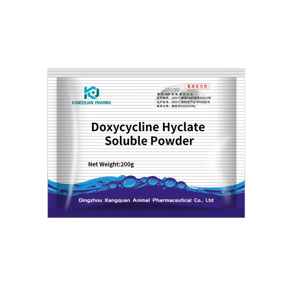
Doxycycline Hyclate Soluble Powder: Your Antibiotic Needs
NewsOct.29,2024
-
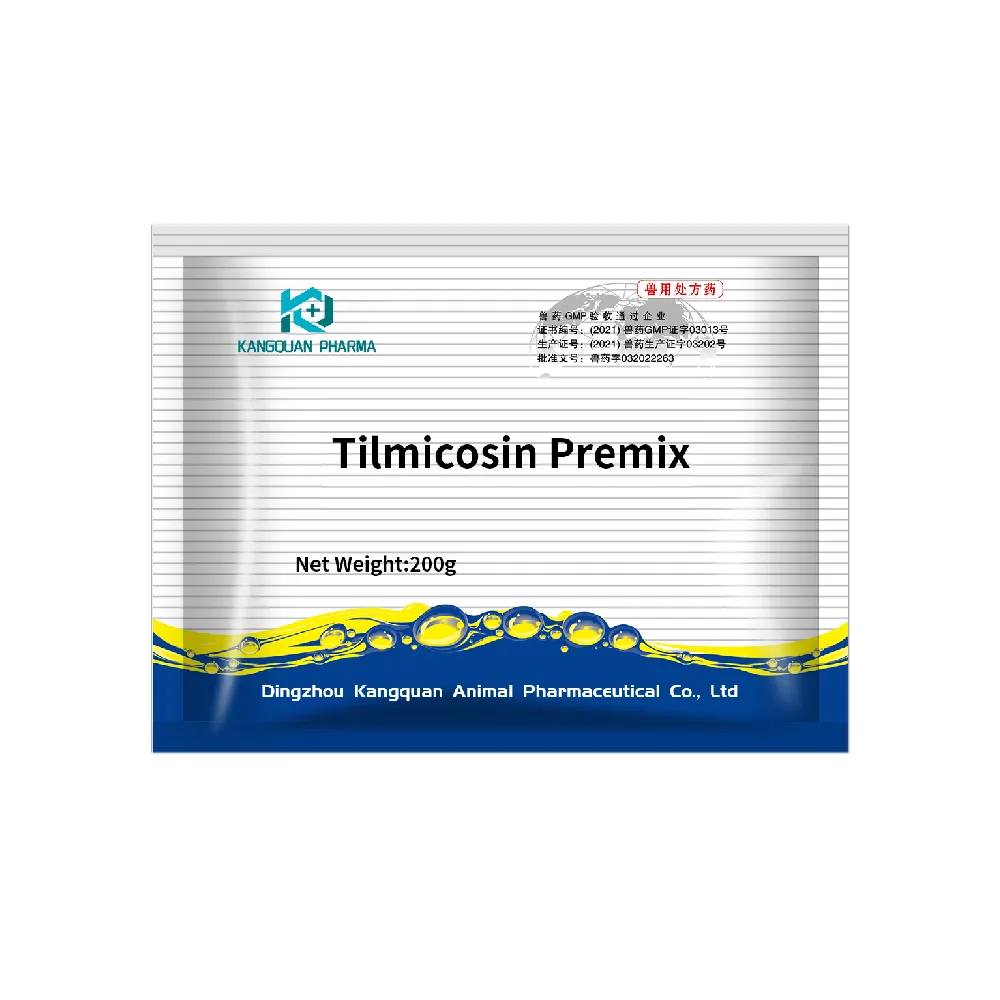
Tilmicosin Premix: The Ultimate Solution for Poultry Health
NewsOct.29,2024
Related Products

Copyright © 2025 Dingzhou Kangquan Pharmaceutical Co., Ltd. All Rights Reserved. Sitemap | Privacy Policy


")