
Info@kangqpharma.com
+86 13780513619
- Afrikaans
- Albanian
- Amharic
- Arabic
- Armenian
- Azerbaijani
- Basque
- Belarusian
- Bengali
- Bosnian
- Bulgarian
- Catalan
- Cebuano
- Corsican
- Croatian
- Czech
- Danish
- Dutch
- English
- Esperanto
- Estonian
- Finnish
- French
- Frisian
- Galician
- Georgian
- German
- Greek
- Gujarati
- Haitian Creole
- hausa
- hawaiian
- Hebrew
- Hindi
- Miao
- Hungarian
- Icelandic
- igbo
- Indonesian
- irish
- Italian
- Japanese
- Javanese
- Kannada
- kazakh
- Khmer
- Rwandese
- Korean
- Kurdish
- Kyrgyz
- Lao
- Latin
- Latvian
- Lithuanian
- Luxembourgish
- Macedonian
- Malgashi
- Malay
- Malayalam
- Maltese
- Maori
- Marathi
- Mongolian
- Myanmar
- Nepali
- Norwegian
- Norwegian
- Occitan
- Pashto
- Persian
- Polish
- Portuguese
- Punjabi
- Romanian
- Russian
- Samoan
- Scottish Gaelic
- Serbian
- Sesotho
- Shona
- Sindhi
- Sinhala
- Slovak
- Slovenian
- Somali
- Spanish
- Sundanese
- Swahili
- Swedish
- Tagalog
- Tajik
- Tamil
- Tatar
- Telugu
- Thai
- Turkish
- Turkmen
- Ukrainian
- Urdu
- Uighur
- Uzbek
- Vietnamese
- Welsh
- Bantu
- Yiddish
- Yoruba
- Zulu
9 月 . 09, 2024 03:55 Back to list
2.5 glutaraldehyde fixation for electron microscopy
2.5% Glutaraldehyde Fixation for Electron Microscopy
Electron microscopy (EM) plays a crucial role in the field of cellular biology, enabling researchers to visualize cellular structures at unprecedented resolution. One of the critical steps in preparing samples for EM is fixation, a process that preserves the cellular architecture by cross-linking proteins and stabilizing membranes. Among the various fixatives used, glutaraldehyde is one of the most effective, particularly at a concentration of 2.5%.
Glutaraldehyde is an aliphatic dialdehyde that reacts with primary amines in proteins, leading to the formation of intra- and intermolecular cross-links. This reaction not only preserves the structural integrity of the cells but also minimizes the diffusion of cellular components, which is crucial for maintaining the natural organization of organelles and membranes. The choice of a 2.5% concentration of glutaraldehyde strikes a balance between effective cross-linking and minimal distortion of cellular structures, making it particularly suitable for high-resolution imaging.
The fixation process with 2.5% glutaraldehyde typically involves several steps. First, the biological sample is immersed in the fixative for a specific duration, often ranging from 1 to 24 hours, depending on the tissue type and desired preservation quality. It is essential to conduct this step under a controlled temperature, usually at 4°C, to slow down cellular metabolism and limit autolysis. Following fixation, samples are washed several times with a buffer, often phosphate-buffered saline (PBS), to remove excess glutaraldehyde, which might interfere with subsequent staining and imaging processes.
2.5 glutaraldehyde fixation for electron microscopy
After fixation and washing, samples are often subjected to additional processing steps, such as osmication, which utilizes osmium tetroxide to further stabilize membranes and enhance contrast in electron microscopy. This dual fixation approach—first with glutaraldehyde and then with osmium—provides optimal preservation of lipid bilayers and protein structures, allowing for detailed ultrastructural analysis.
The use of 2.5% glutaraldehyde fixation has been widely adopted in diverse fields of biological research, including neurobiology, pathology, and cell biology. The technique has enabled significant advancements in our understanding of cellular processes, organelle dynamics, and pathological changes associated with diseases.
In conclusion, the fixation of biological samples with 2.5% glutaraldehyde is a pivotal step in preparing specimens for electron microscopy. Its ability to effectively preserve cellular structures while allowing for high-resolution imaging makes it a favored choice in various scientific applications. As electron microscopy continues to evolve, the importance of proper fixation techniques will remain paramount in unlocking the mysteries of cell biology.
Latest news
-
The Power of Radix Isatidis Extract for Your Health and Wellness
NewsOct.29,2024
-
Neomycin Sulfate Soluble Powder: A Versatile Solution for Pet Health
NewsOct.29,2024
-
Lincomycin Hydrochloride Soluble Powder – The Essential Solution
NewsOct.29,2024
-
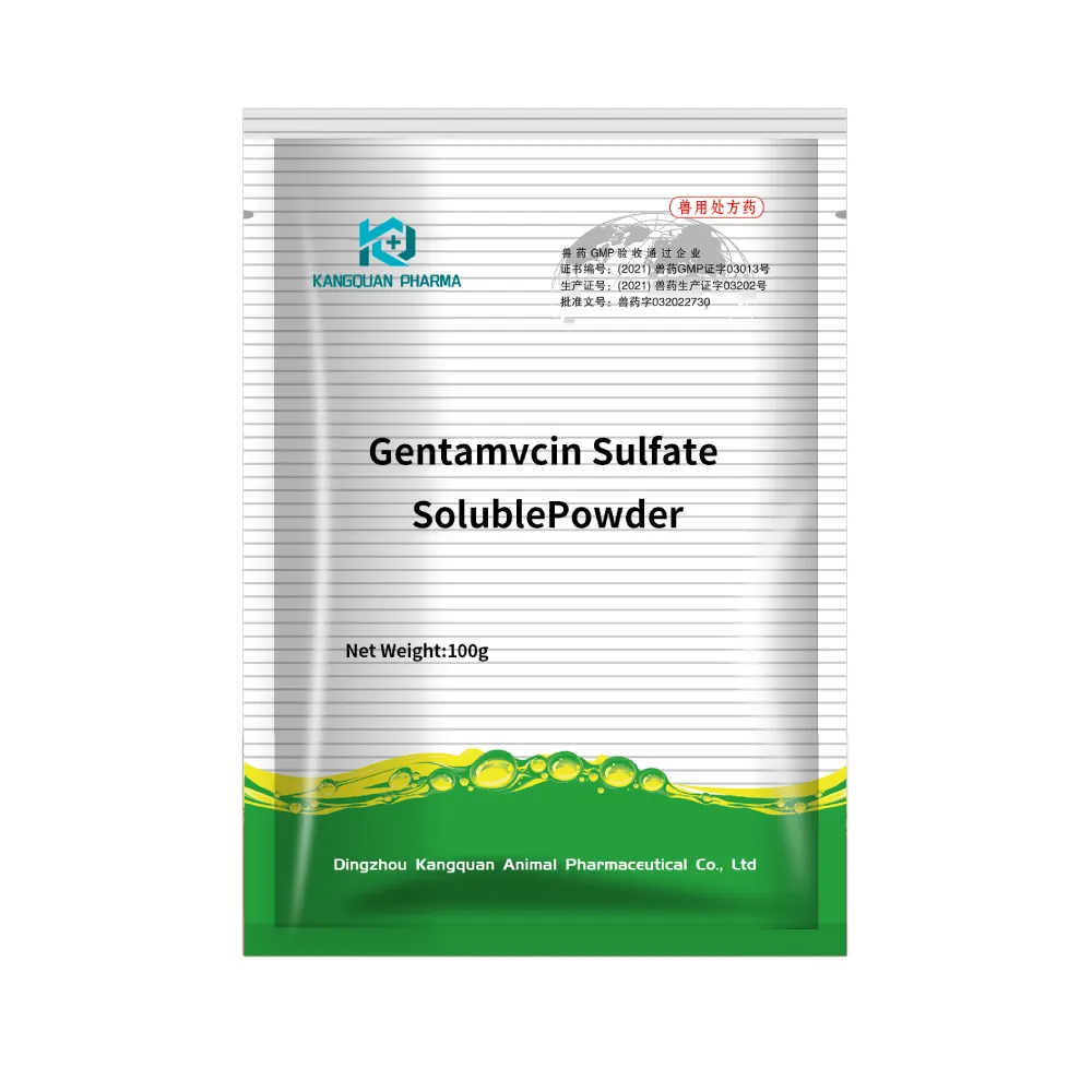
Garamycin Gentamicin Sulfate for Effective Infection Control
NewsOct.29,2024
-
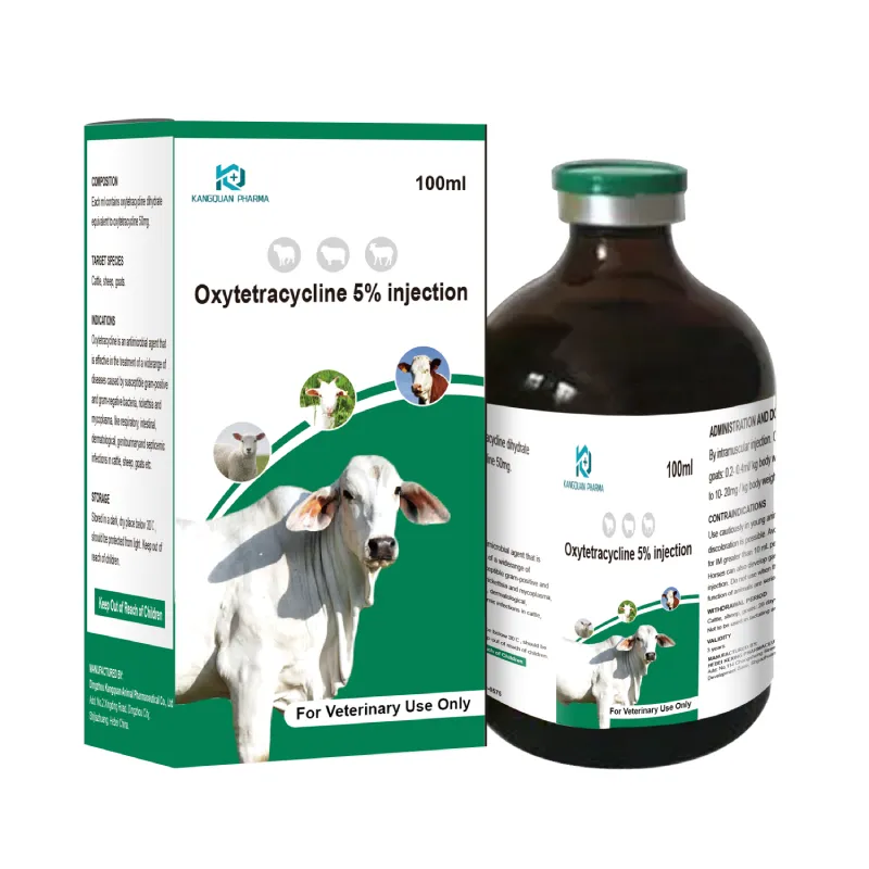
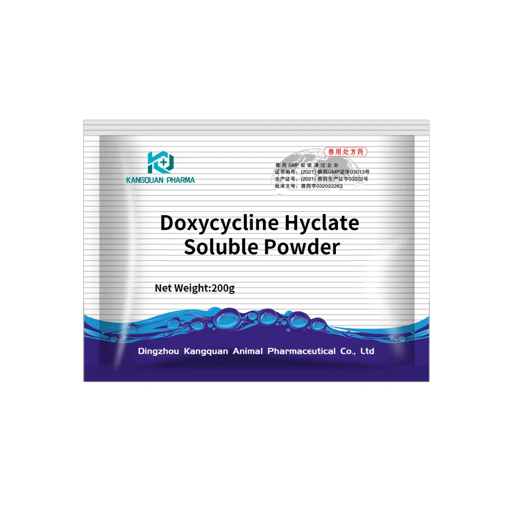
Doxycycline Hyclate Soluble Powder: Your Antibiotic Needs
NewsOct.29,2024
-
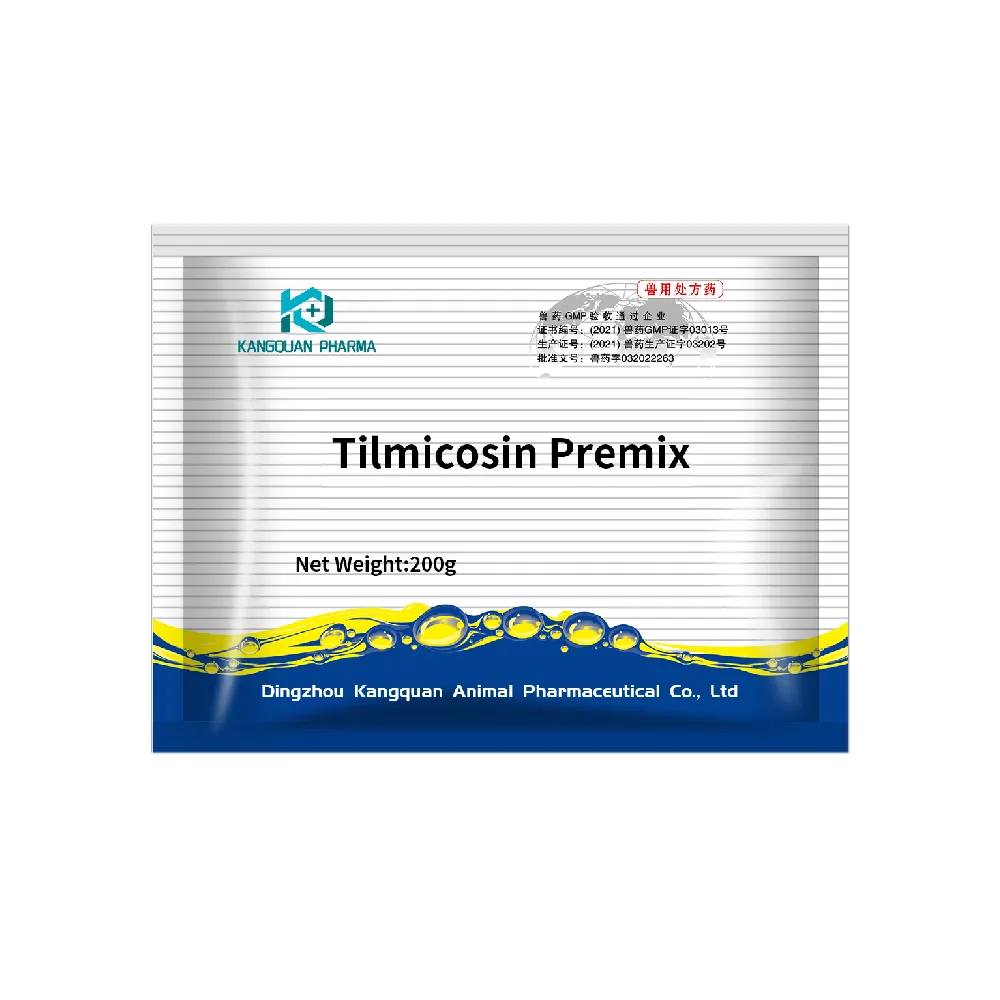
Tilmicosin Premix: The Ultimate Solution for Poultry Health
NewsOct.29,2024
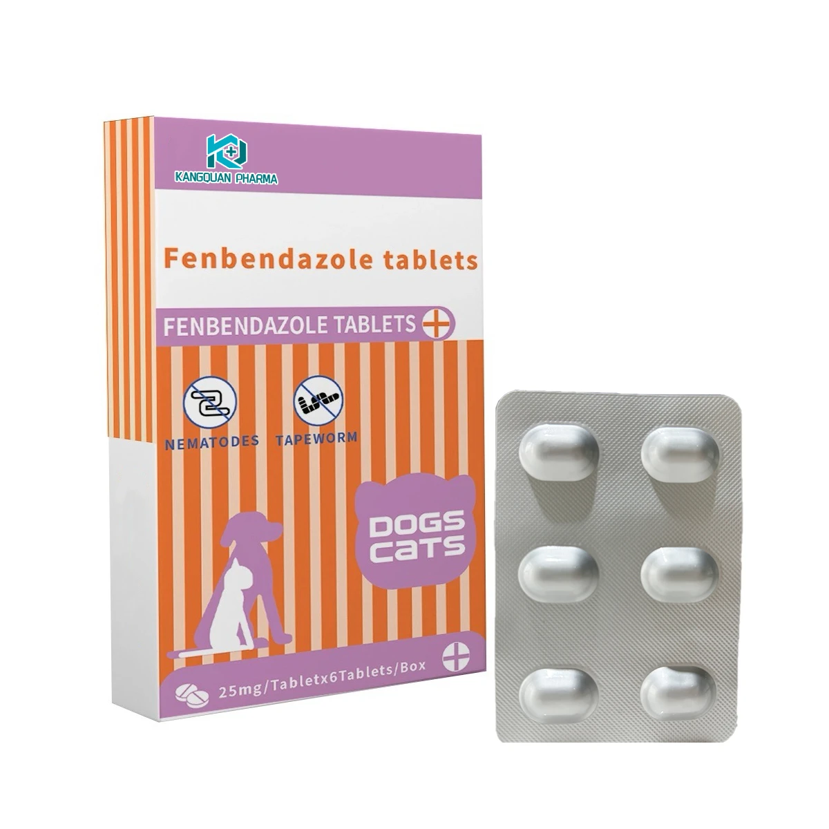
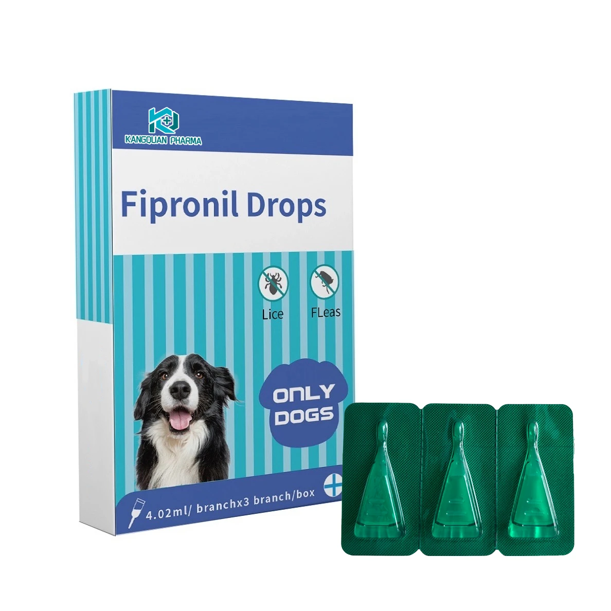
Related Products

Copyright © 2025 Dingzhou Kangquan Pharmaceutical Co., Ltd. All Rights Reserved. Sitemap | Privacy Policy


")